

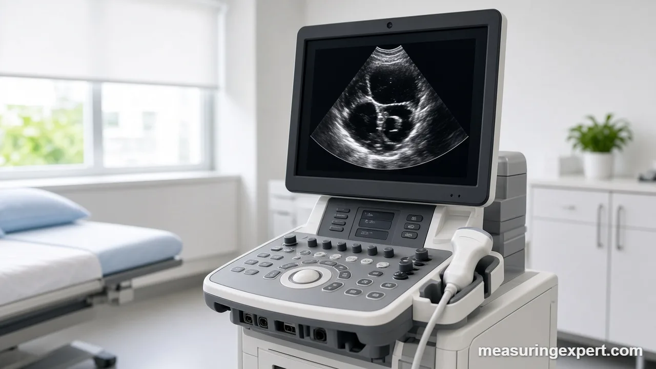
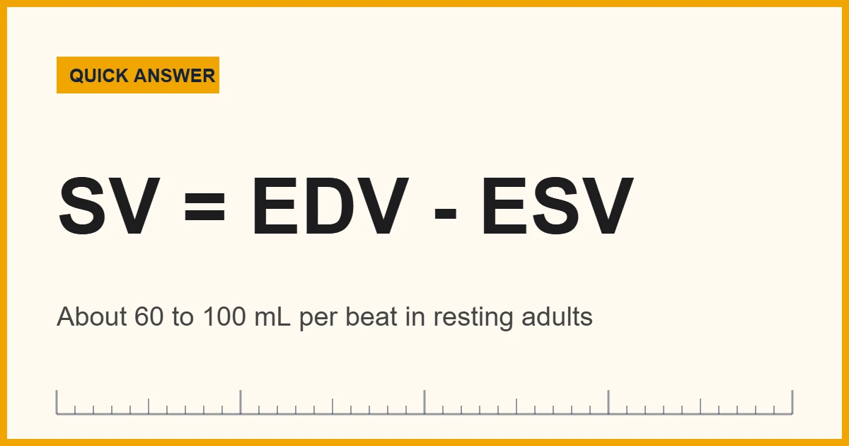
Stroke volume is the amount of blood one ventricle pushes out with each heartbeat. Doctors usually measure it with an echocardiogram, where SV = EDV – ESV. In healthy adults at rest, it runs about 60 to 100 mL (2 to 3.4 US fl oz) per beat, and cardiac output equals SV times heart rate.
In fact, your heart does not pump a fixed amount. Instead, every beat fills the main pumping chamber, the left ventricle, and then squeezes out part of that blood into the aorta. The part that leaves is the stroke volume. As a result, it is one of the most useful single numbers in heart physiology: it shows how well the heart fills, how hard it squeezes, and how much blood the body actually receives.
In short: you cannot measure stroke volume at home with a cuff, a watch or a pulse oximeter. Instead, clinicians estimate it from images of the heart (echocardiography or cardiac MRI), from blood-flow speed measured with Doppler ultrasound, or from invasive catheter methods in hospital settings.
The US National Library of Medicine’s StatPearls review Physiology, Stroke Volume defines it as the difference between end-diastolic volume and end-systolic volume, and it gives a normal resting range of 60 to 100 mL in healthy adults. Similarly, its companion review on cardiac output explains that cardiac output is SV multiplied by heart rate.
This guide is educational. First, it walks through the formulas with worked numbers. Next, it compares the main measurement methods and explains what the results mean. Finally, it covers the honest limits of each approach and when a number on a report deserves a conversation with your doctor.
Convert a Stroke Volume Reading to Other Units
Echo reports list stroke volume in milliliters. So, if you want to picture that amount in liters, fluid ounces or teaspoons, type the value below. The default of 70 mL is a typical resting adult figure, not a target.
For context, a 70 mL beat is about 2.37 US fluid ounces, or roughly 14 teaspoons. To turn it into cardiac output, multiply by your heart rate; the formulas section below shows how.
Recommended Tools for Tracking Heart Rate and Blood Pressure at Home
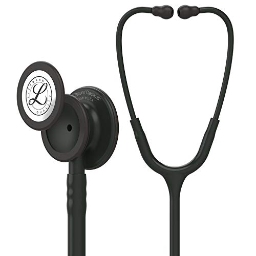
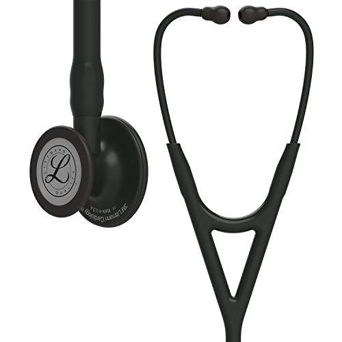
No consumer device measures stroke volume directly. However, the other half of the cardiac output equation, heart rate, is easy to track at home, and blood pressure readings give your doctor useful context. For example, a validated upper-arm blood pressure monitor, a chest-strap heart rate sensor and a good stethoscope cover most home and student needs. Even so, treat these as tracking tools, not diagnostic tests.


As an Amazon Associate, Measuring Expert earns from qualifying purchases.
- Stroke volume (SV) is the blood ejected by one ventricle in one beat.
- The core formula is SV = end-diastolic volume (EDV) minus end-systolic volume (ESV).
- Healthy adults at rest usually fall between 60 and 100 mL per beat.
- Cardiac output = SV x heart rate; at rest it is typically about 5 to 6 L/min.
- Echocardiography is the everyday clinical method; cardiac MRI is more precise for volumes.
- Doppler echo can also estimate SV from the outflow tract area and blood-flow speed.
- Only a clinician can judge whether your number is normal for you.
Stroke Volume Formulas: SV, Cardiac Output and Ejection Fraction
Three short equations explain almost everything on an echo report. Once you see how they connect, the numbers stop looking random.
SV = EDV – ESVEnd-diastolic volume (EDV) is how much blood the left ventricle holds when it is fullest, just before it contracts. By contrast, end-systolic volume (ESV) is what stays behind after it contracts. For example, a ventricle that fills to 120 mL and keeps 50 mL ejects 70 mL. Therefore, that 70 mL is the stroke volume.
Cardiac output (L/min) = SV (mL) x heart rate (beats/min) / 1,000Next, multiply by heart rate. With 70 mL per beat and 70 beats per minute, the heart moves 4,900 mL, or 4.9 L, every minute. Because heart rate is part of the equation, it helps to know your true resting pulse; our guide on the best time to measure resting heart rate explains how to get a clean reading.
Stroke volume index and ejection fraction
Ejection fraction (%) = SV / EDV x 100Ejection fraction (EF) expresses the same beat as a percentage. Using the example above, 70 / 120 x 100 gives about 58%. In other words, the ventricle pushed out a little over half of the blood it held. Doctors also sometimes divide SV by body surface area to get the SV index, so that a tall adult and a small adult can be compared fairly; 70 mL in a person with 1.9 square meters of body surface gives about 36.8 mL/m2.
| Term | What it means | Worked example |
|---|---|---|
| EDV | Volume in the ventricle when fullest | 120 mL |
| ESV | Volume left after contraction | 50 mL |
| Stroke volume (SV) | EDV – ESV, blood ejected per beat | 70 mL |
| Heart rate (HR) | Beats per minute | 70 bpm |
| Cardiac output (CO) | SV x HR | 4.9 L/min |
| Ejection fraction (EF) | SV / EDV | about 58% |
| SV index | SV / body surface area | about 36.8 mL/m2 (at 1.9 m2) |
Normal Stroke Volume Ranges in Adults
A single “normal” number does not exist, because body size, age, sex, fitness and posture all shift it. Still, reference ranges give a useful frame. However, the figures below describe healthy adults at rest; they are not targets, and they are not a way to judge your own report.
| Measure | Typical resting adult value | Source |
|---|---|---|
| Stroke volume | 60 to 100 mL per beat (about 2.0 to 3.4 US fl oz) | StatPearls, Physiology, Stroke Volume |
| Cardiac output | About 5 to 6 L/min at rest; over 35 L/min in elite athletes during exercise | StatPearls, Physiology, Cardiac Output |
| Left ventricular ejection fraction | Roughly 52% to 74% in adults by echo, with slightly different cutoffs for men and women | American Society of Echocardiography |
| Resting heart rate | Commonly quoted as 60 to 100 bpm for adults | General clinical reference |
How Stroke Volume Is Measured: Methods Compared
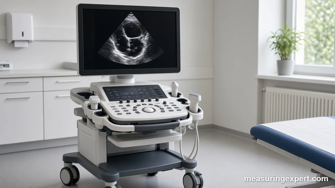
Several techniques can estimate it. They differ in how invasive they are, where they are used and what they actually measure. However, most people meet only the first one, the echocardiogram.
| Method | How it works | Invasive? | Typical setting |
|---|---|---|---|
| 2D echocardiography (biplane method of discs) | Traces the ventricle outline at end-diastole and end-systole, then calculates EDV and ESV | No | Clinic, hospital |
| Doppler echocardiography (LVOT VTI) | Multiplies outflow tract area by the distance blood travels per beat | No | Clinic, ICU, emergency |
| 3D echocardiography | Measures the ventricle volume directly from a 3D data set | No | Specialist echo labs |
| Cardiac MRI | Stacks short-axis slices to measure volumes very precisely | No | Imaging centers |
| Thermodilution | Injects cool fluid through a pulmonary artery catheter and tracks the temperature change | Yes | ICU, cath lab |
| Fick principle | Calculates cardiac output from oxygen uptake and blood oxygen content, then divides by heart rate | Yes (direct Fick) | Cath lab, research |
| Impedance cardiography and pulse contour devices | Estimate flow from electrical signals or the arterial pressure waveform | Partly | Monitoring, research |
Each method has a niche. For example, cardiac MRI is often treated as the reference for chamber volumes, while Doppler echo is quick enough to repeat at the bedside every few minutes. Meanwhile, invasive catheter methods are reserved for very sick patients who already need monitoring lines.
How an Echocardiogram Measures Stroke Volume
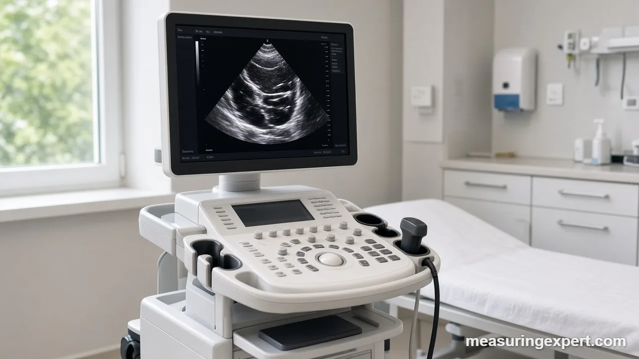
An echocardiogram is an ultrasound scan of the heart. You lie on your left side, a sonographer puts gel on your chest, and a handheld probe sends sound waves that bounce back as moving images. Also, the scan is painless and uses no radiation. From those images, the software and the reader can estimate SV in two main ways.
The volume method (Simpson’s biplane)
First, the sonographer records two standard views of the left ventricle. Then the inner border of the chamber is traced at its largest (end-diastole) and smallest (end-systole) moments. Next, the software slices each outline into a stack of thin discs and adds their volumes. As a result, you get EDV and ESV, and the difference is the stroke volume. The same tracing gives ejection fraction.
The Doppler method (LVOT VTI)
Instead of chamber size, this method measures flow. The sonographer first measures the diameter of the left ventricular outflow tract (LVOT), the short channel blood passes through on its way to the aorta. Next, Doppler records the speed of blood there, and the machine integrates it over one beat to give the velocity time integral (VTI), in centimeters. In other words, think of it as a cylinder of blood:
SV (mL) = 0.785 x LVOT diameter (cm)^2 x VTI (cm)For example, a 2.0 cm outflow tract and a VTI of 20 cm give 0.785 x 4 x 20, or about 62.8 mL. Because the diameter is squared, a small measuring error matters a lot. For instance, a 2.2 cm diameter with the same VTI gives about 76.0 mL, which is roughly 21% more.
How to Calculate Stroke Volume From Echo Numbers
If you are a student or simply curious, you can reproduce the arithmetic from the values on a report. However, use this only for learning; your cardiologist’s interpretation is what counts.
- Find EDV and ESV. Look for “LV EDV” and “LV ESV” (often from the biplane method), both in mL.
- Subtract. Take EDV minus ESV; for example, 130 – 55 = 75 mL.
- Check ejection fraction. Divide your result by EDV and multiply by 100; here, 75 / 130 gives about 58%.
- Add the heart rate. Use the heart rate recorded during the scan, not a reading from another day.
- Work out cardiac output. Multiply mL by beats per minute and divide by 1,000; for instance, 75 x 62 / 1,000 = 4.65 L/min.
- Compare with the report. If your number differs, the lab may have used Doppler or 3D data instead.
What Changes the Output of Each Beat
Three physiological levers set SV, and doctors think about all three when they read the number.
- Preload. This is how much the ventricle fills. More blood returning to the heart stretches the muscle, so it contracts more strongly (the Frank-Starling mechanism). Dehydration or blood loss lowers it.
- Contractility. This is how forcefully the muscle squeezes. Adrenaline raises it, while a damaged heart muscle lowers it.
- Afterload. This is the resistance the ventricle pushes against. High blood pressure or a narrowed aortic valve makes ejection harder.
Besides heart function, other everyday factors matter too. For example, standing up briefly reduces the blood returning to the heart, and exercise increases both SV and heart rate. Similarly, a very fast heart rate can leave too little time for filling, so each beat carries less. Lung function interacts with all of this as well; our explainer on why residual lung volume cannot be measured directly shows a similar “measure it indirectly” problem in the lungs.
Reading Echo Results: Do and Don’t
Do
- Ask which method the lab used for the stroke volume figure.
- Compare reports from the same lab and method when tracking change.
- Note your heart rate and blood pressure on the day of the scan.
- Bring questions about any flagged value to your doctor.
Don’t
- Treat a wearable’s “cardiac output” estimate as a medical measurement.
- Change medicines based on a number you calculated yourself.
- Assume one value outside a textbook range means disease.
- Ignore symptoms because a past report looked normal.
Honest Limits of Stroke Volume Measurement
Every method estimates SV rather than weighing the blood directly. For one thing, 2D echo assumes the ventricle has a regular shape, so it tends to underestimate volumes compared with cardiac MRI. In addition, image quality depends on body build, lung disease and the sonographer’s skill. Similarly, with Doppler, a 1 to 2 millimeter error in the outflow tract diameter changes the result noticeably, because that value is squared. Moreover, an irregular rhythm such as atrial fibrillation makes every beat different, so labs average several beats. Finally, invasive methods are accurate in the right hands but carry risks that make sense only in hospital care. In short, treat any single SV number as an estimate with a margin of error, and focus on trends and the overall clinical picture.
When to Talk to a Doctor
Ask your doctor or cardiologist to explain your report if it flags a low ejection fraction, a low SV or cardiac output, or enlarged chamber volumes. Also ask if you have symptoms such as breathlessness on exertion, swollen ankles, unusual fatigue or palpitations. After that, a doctor can combine the echo with your history, an ECG, blood tests and, if needed, cardiac MRI or catheter studies. However, this page cannot do that, and no home device can either.
Stroke Volume FAQs
What is stroke volume in simple terms?
It is the amount of blood one ventricle pushes out with a single heartbeat, usually reported in milliliters for the left ventricle.
What is the formula for stroke volume?
SV = EDV – ESV: end-diastolic volume minus end-systolic volume. Doppler echo can also estimate it as 0.785 x LVOT diameter squared x VTI.
What is a normal stroke volume?
Healthy adults at rest usually fall between about 60 and 100 mL per beat. However, size, age, sex and fitness all shift the value.
How do you calculate cardiac output from stroke volume?
Multiply SV in mL by heart rate in beats per minute, then divide by 1,000. For example, 70 mL x 70 bpm = 4.9 L/min.
Which test measures stroke volume?
An echocardiogram is the most common test. Cardiac MRI, thermodilution catheters and the Fick method are also used in specific situations.
Can a smartwatch measure stroke volume?
No. Watches and fitness bands measure heart rate. Any SV or cardiac output figure they show is a modeled estimate, not a clinical measurement.
Is stroke volume the same as ejection fraction?
Not quite. Stroke volume is a volume in mL, while ejection fraction is that volume divided by end-diastolic volume, shown as a percentage.
Why do athletes pump more blood per beat?
Endurance training enlarges and strengthens the heart, so each beat ejects more blood. That is why many athletes have a slow resting pulse.
What makes each beat pump less blood?
Dehydration, blood loss, a weakened heart muscle, valve narrowing, high blood pressure and very fast heart rates can all reduce it.
Should I worry about a value outside the normal range?
Ask your doctor. A single value needs context from your symptoms, other measurements and history before anyone can say what it means.
The Bottom Line on Measuring Stroke Volume
To sum up, stroke volume is the blood the heart ejects per beat, calculated as EDV minus ESV and usually about 60 to 100 mL in resting adults. Multiply it by heart rate and you get cardiac output, typically about 5 to 6 L/min at rest. Echocardiography is the everyday way to measure it, with Doppler and cardiac MRI adding detail.
Above all, treat these numbers as tools for understanding, not self-diagnosis. If a report or a symptom worries you, the right next step is a conversation with your doctor.
1 thought on “How Is Stroke Volume Measured? Echo, Formulas and Normal Ranges”