

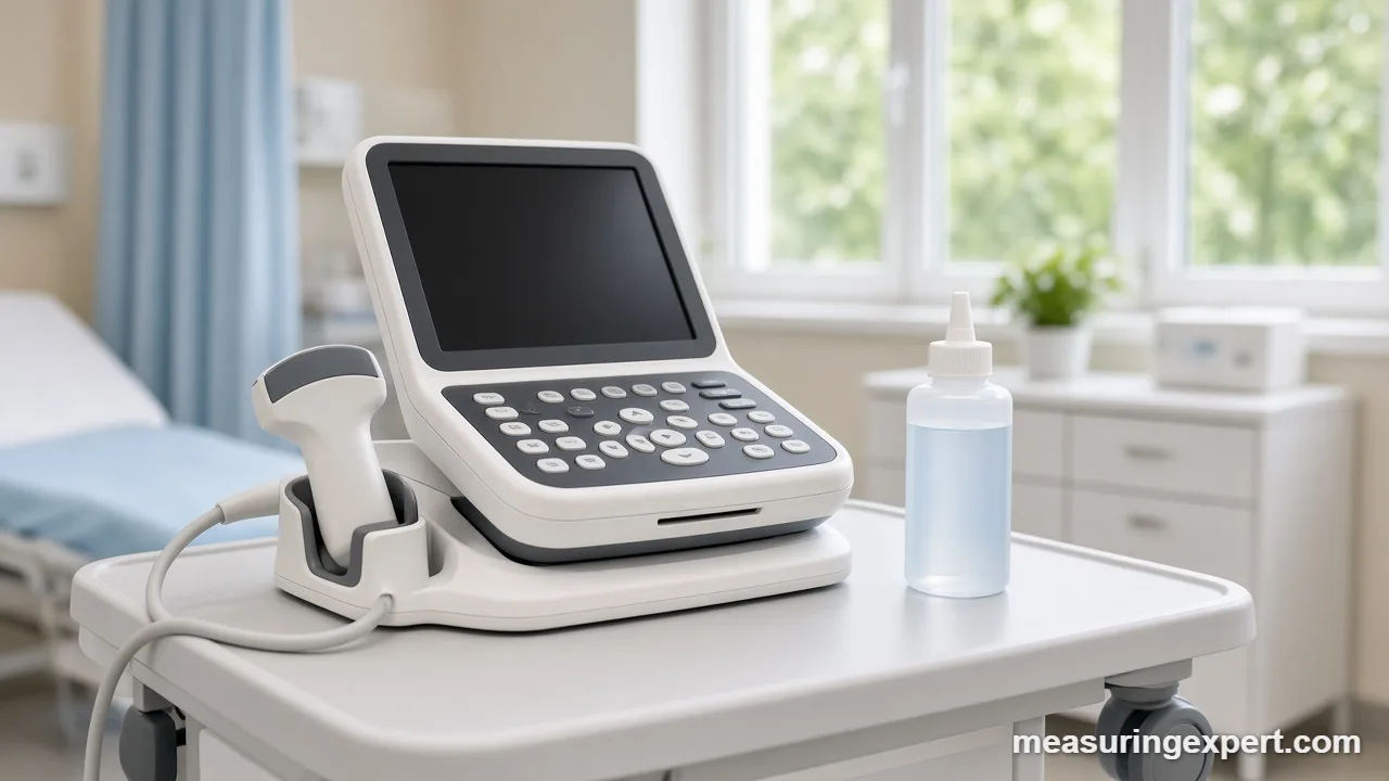
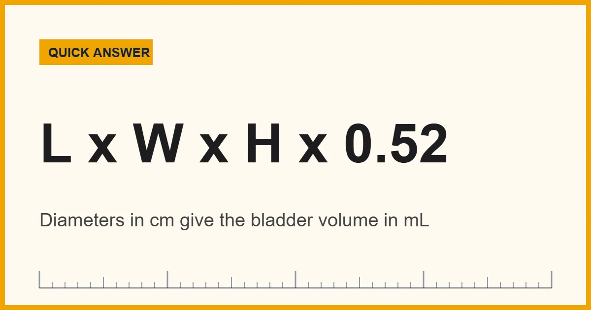
Bladder volume on ultrasound is usually estimated with the ellipsoid formula: volume (mL) = length x width x height (cm) x 0.52. So a bladder measuring 8 x 6 x 5 cm holds about 125 mL. However, the 0.52 coefficient is a convention, and published studies suggest values up to about 0.89 depending on bladder shape.
In particular, this guide is for students, sonographers, nurses and clinicians who want to understand how the number on the report is produced. First, it walks through the ellipsoid formula and where the 0.52 comes from. Next, it covers post-void residual (PVR) volume, the measurement most bladder scans are ordered for. Finally, it compares manual 2D measurement with automated bladder scanners and catheter drainage.
In short: you take three perpendicular diameters of the urine-filled bladder in centimeters, multiply them, and then multiply by a correction coefficient. Because 1 cubic centimeter equals 1 milliliter, the result is already in mL.
The method is simple, so the hard part is measuring well. For example, a probe that sits slightly off the true midline shortens a diameter, and a small error in each of three numbers multiplies into a larger error in the volume.
StatPearls, the peer-reviewed clinical reference hosted by the US National Library of Medicine, describes the same length x width x height x 0.52 calculation for post-void residual volume. It also lists the adult PVR ranges that clinicians commonly use, which appear in a table further down.
Convert a Bladder Volume From mL to Other Units
First, multiply your three diameters and the coefficient to get milliliters. Then enter that mL value below to see it in cubic centimeters, liters, US fluid ounces and cups. As a result, you can check a worked example or a textbook question in whichever unit it uses.
Recommended Supplies for Learning Bladder Scanning
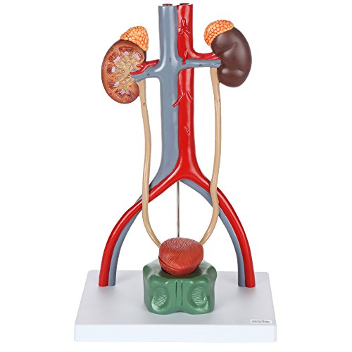
Hospital bladder scanners and diagnostic ultrasound systems are medical devices bought through clinical suppliers, so they are not listed here. Instead, these items help students practice and study. For instance, clear coupling gel suits training scans, an anatomy model helps with orientation, and a graduated cylinder shows what a given volume of fluid really looks like.




As an Amazon Associate, Measuring Expert earns from qualifying purchases.
Key Takeaways
- Standard estimate: volume (mL) = L x W x H (cm) x 0.52.
- The 0.52 comes from the ellipsoid volume formula, pi/6, which is about 0.5236.
- Real bladders are rarely perfect ellipsoids, so one study found 0.72 a better average coefficient.
- Post-void residual (PVR) means the urine left in the bladder right after voiding.
- Adult PVR under about 100 mL is commonly treated as normal.
- Automated bladder scanners give a 3D estimate in under a minute, while catheter drainage remains the reference standard.
- Any single number needs clinical context; one reading does not make a diagnosis.
The Bladder Volume Ellipsoid Formula Explained
In general, a full bladder looks roughly like a rounded box or an egg on ultrasound. So instead of tracing its exact outline, most protocols treat it as an ellipsoid, a stretched sphere with three axes. The volume of an ellipsoid equals pi/6 multiplied by its three full diameters, and pi/6 works out to about 0.5236. Therefore, rounded to two decimals, that gives the familiar formula:
Volume (mL) = length (cm) x width (cm) x height (cm) x 0.52
Each dimension, however, comes from a different view. First, the transverse view across the lower abdomen gives the width (side to side). Next, the sagittal (longitudinal) view gives the length (head to foot). Finally, the height, also called depth or anteroposterior (AP) diameter, runs front to back and can come from either view. In any case, sonographers take the largest value of each.
In addition, the units work out neatly. Because centimeters multiplied three times give cubic centimeters, and 1 cc equals exactly 1 mL, the answer needs no further conversion. If you ever have the diameters in millimeters, however, divide the final product by 1,000.
Of course, the same idea, measuring shape and applying a geometric formula, appears across measurement science. For a non-medical comparison, see how to find the volume of an irregular object by water displacement, which avoids shape assumptions entirely.
Why the Bladder Volume Coefficient Varies
Specifically, the 0.52 coefficient assumes a perfect ellipsoid. Real bladders, however, change shape as they fill, and they look different in children, after pelvic surgery, or next to an enlarged prostate or pregnant uterus. As a result, researchers have tested other coefficients.
In one widely cited study, Bih and colleagues compared ten formulas against catheter volumes in 24 healthy volunteers and 55 people with spinal cord injury. They then grouped bladders by shape and found that the best general coefficient was 0.72. In addition, shape-specific coefficients cut the mean error further.
| Bladder shape (Bih et al., 1998) | Best-fit coefficient | Volume for 8 x 6 x 5 cm |
|---|---|---|
| Textbook ellipsoid (pi/6) | 0.52 | 124.8 mL |
| Triangular prism | 0.66 | 158.4 mL |
| All shapes combined | 0.72 | 172.8 mL |
| Ellipsoid (as measured in the study) | 0.81 | 194.4 mL |
| Cuboidal | 0.89 | 213.6 mL |
The same three diameters can therefore produce estimates from about 125 to 214 mL. For this reason, reports should state the coefficient. Likewise, trends over time on the same machine and protocol mean more than one isolated value.
How to Measure Bladder Volume on Ultrasound
The steps below describe the general technique taught in training. However, local protocols, machine software and supervision rules always come first.
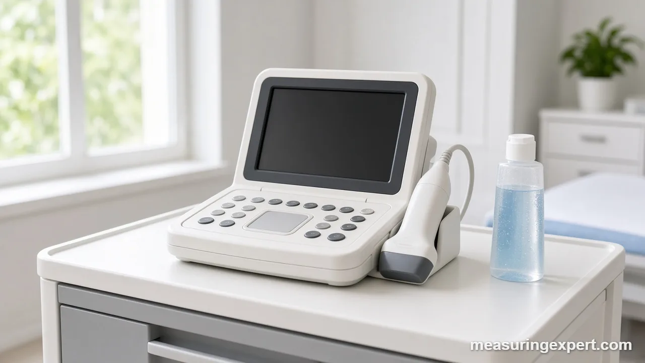
- Prepare the patient and machine. Explain the scan, position the patient supine, and select a curvilinear (abdominal) probe with a bladder or pelvic preset.
- Apply gel and find the bladder. Place the probe just above the pubic bone, angle it slightly toward the feet, and look for the dark, fluid-filled bladder.
- Take the transverse view. With the probe marker toward the patient’s right, freeze the image where the bladder looks widest and measure its width.
- Measure height in the same view. Then place calipers on the inner walls from front to back for the AP diameter.
- Rotate to the sagittal view. Turn the probe 90 degrees, marker toward the head, and measure the longest length from top to bottom.
- Calculate the volume. Multiply length x width x height in centimeters by your coefficient, or let the machine’s bladder package do it.
- Record and repeat if needed. Note the dimensions, coefficient, timing relative to voiding, and the result in mL; repeat once if the image quality was poor.
Bladder Volume Worked Examples
These examples use the standard 0.52 coefficient, with the 0.72 value alongside for comparison. Also, each product is in cubic centimeters, which equals mL.
| Length x width x height (cm) | Product (cc) | x 0.52 | x 0.72 |
|---|---|---|---|
| 4 x 3 x 2.5 | 30 | 15.6 mL | 21.6 mL |
| 8 x 6 x 5 | 240 | 124.8 mL | 172.8 mL |
| 10 x 9 x 7 | 630 | 327.6 mL | 453.6 mL |
| 12 x 10 x 8 | 960 | 499.2 mL | 691.2 mL |
For instance, take the second row. First, 8 x 6 = 48. Next, 48 x 5 = 240 cc. Finally, 240 x 0.52 = 124.8 mL, which is about 4.22 US fluid ounces. Similarly, the third row gives 327.6 mL, so the same arithmetic scales to any size.
Above all, notice how sensitive the result is. If each diameter in the 8 x 6 x 5 example reads just 0.5 cm long, the product becomes 8.5 x 6.5 x 5.5, and the estimate rises from about 125 to about 158 mL.
Post-Void Residual: What the Bladder Volume Means
Post-void residual (PVR) is the amount of urine left in the bladder right after a person urinates. Clinicians measure it, for example, when someone has trouble emptying, after surgery or catheter removal, or with certain nerve conditions. Ideally, the scan happens within a few minutes of voiding, because the kidneys keep adding urine.
StatPearls gives the following commonly used adult ranges. Even so, cutoffs differ between guidelines, age groups and clinical settings, so they are a general reference rather than a rule.
| Adult PVR | Common interpretation (StatPearls) | US fluid ounces |
|---|---|---|
| Under 100 mL | Normal | under 3.4 fl oz |
| 100 to 200 mL | Acceptable | 3.4 to 6.8 fl oz |
| Over 200 mL | Inadequate emptying | over 6.8 fl oz |
| Over 300 mL | Suggests urinary retention | over 10.1 fl oz |
| Over 400 mL | Diagnostic of urinary retention | over 13.5 fl oz |
Bladder Volume by Scanner, Ultrasound or Catheter
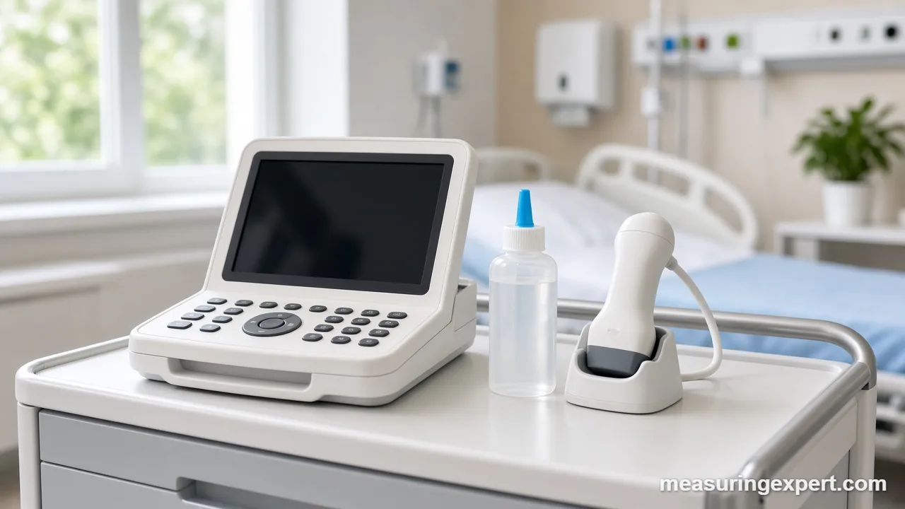
In general, there are three common ways to get a bladder volume, and each trades convenience against accuracy.
| Method | How it works | Strengths | Drawbacks |
|---|---|---|---|
| Automated bladder scanner | A handheld ultrasound probe sweeps several planes and software estimates a 3D volume. | Fast (StatPearls cites about 45 seconds), noninvasive, nurse-friendly. | Readings can be thrown off by ascites, cysts, obesity, pregnancy or poor probe aim. |
| Formal 2D ultrasound | A sonographer measures three diameters and applies the ellipsoid formula. | Shows the anatomy, so it can also spot other problems. | Needs trained staff; the shape assumption adds error. |
| Catheter drainage | A catheter drains the bladder into a graduated container. | Reference standard for volume. | Invasive, slower, and carries infection and trauma risk. |
Because scanners are quick and noninvasive, many wards use them for routine PVR checks. However, StatPearls notes that accuracy data are mixed, so a surprising reading usually prompts a repeat scan or a different method.
Similarly, volume estimates from shape measurements show up elsewhere in medicine. Our guide on how to measure stroke volume covers how echocardiography estimates the blood the heart pumps per beat.
Bladder Volume Do and Don’t List
Do
- Measure all three diameters at their largest.
- Place calipers on the inner wall.
- Record the coefficient with the result.
- Scan soon after voiding for PVR.
- Repeat a reading that looks out of place.
Don’t
- Mix millimeters and centimeters in one calculation.
- Compare volumes made with different coefficients.
- Rely on one reading for a clinical decision.
- Assume every dark pelvic structure is bladder.
- Interpret scans outside your training or role.
Honest Limits of Ultrasound Estimates
An ultrasound bladder volume is an estimate, not a direct measurement. First, the formula assumes a shape the bladder rarely has, and the Bih study shows that this alone can shift results by more than half. Second, small caliper errors multiply across three diameters. Third, operator technique, patient position and body habitus all matter, and fluid collections near the bladder can mislead automated scanners.
In addition, very small volumes are harder to measure well, because the walls are close together and a millimeter or two becomes a large share of each diameter. So treat the number as a useful, fast estimate. When precision really matters, clinicians confirm it with a repeat scan or catheter drainage.
When to Involve a Professional
Learning the formula is one thing; performing and interpreting scans is another. Therefore, students should scan only under supervision and within their program’s rules. Likewise, nurses and clinicians follow their facility’s bladder scanning policy and escalate unexpected readings.
If you are a patient, or you are reading about a family member, please do not try to work out a diagnosis from these numbers. Instead, ask the doctor or nurse who ordered the scan what the result means. Also, seek urgent medical help for sudden inability to urinate, severe lower abdominal pain, or fever with urinary symptoms.
Frequently Asked Questions
What is the formula for bladder volume on ultrasound?
The usual formula is volume (mL) = length x width x height in centimeters x 0.52. Specifically, the three diameters come from transverse and sagittal views of the full bladder.
Why is 0.52 used?
The volume of an ellipsoid is pi/6 times its three diameters, and pi/6 is about 0.5236. As a result, rounding gives 0.52.
Is 0.52 always the right bladder volume coefficient?
No. Bladders are not perfect ellipsoids, and one study found 0.72 a better general coefficient, with shape-specific values from 0.66 to 0.89. So use the value your protocol specifies.
Are cc and mL the same for bladder volume?
Yes. One cubic centimeter equals exactly one milliliter, so a result in cc is the same number in mL.
What is post-void residual?
Post-void residual (PVR) is the urine left in the bladder right after urinating. Clinicians usually measure it with a bladder scanner, formal ultrasound or a catheter.
What PVR is considered normal?
StatPearls lists adult PVR under 100 mL as normal and 100 to 200 mL as acceptable. However, cutoffs vary by guideline and patient group, and a clinician interprets each result.
How accurate are bladder scanners?
They are fast and noninvasive, but studies show mixed accuracy. Ascites, cysts, obesity, pregnancy and poor probe aim can distort readings, so unexpected values are usually rechecked.
How do I convert bladder volume from mL to ounces?
Divide mL by 29.57 to get US fluid ounces. For example, 125 mL is about 4.2 fl oz, and the converter above does this for any value.
Which probe is used to measure bladder volume?
Transabdominal scans typically use a low-frequency curvilinear probe. By contrast, dedicated bladder scanners have their own built-in probe and software.
Can I interpret my own bladder scan result?
No. This article is educational only. Instead, ask the doctor or nurse who ordered the scan to explain your result.
Bladder Volume on Ultrasound: The Bottom Line
To sum up, ultrasound estimates bladder volume by multiplying three perpendicular diameters in centimeters by a coefficient, most often 0.52. The math is easy, but shape, caliper placement and the chosen coefficient can change the answer a lot.
So measure carefully, record your method, and treat every number as an estimate that a qualified clinician interprets. Finally, for quick unit checks, use the converter near the top of this page.